External Injuries to Face and Scalp – Dr Andrew has several decades of experience examining and interpreting head injuries. Below he outlines the many forms that head injuries can take.
- Abrasions – tangential application of force resulting in scraping of superficial layers of skin
- Lacerations – direct force applied to a more defined area of enough magnitude to tear the skin. May be partial or full thickness
- Undermining – lifting of the lacerated skin off the underlying tissues or bone, with formation of a pocket. Indicates direction of applied force
- Scalp Hemorrhage – a type of closed head injury with no breach of the skin
- Subscalpular – bleeding in the subcutaneous tissues of the scalp
- Subgaleal – bleeding beneath the protective periosteal layer of the skull (galea)
- Skull Fracture
- Open v. closed
- Complete v. incomplete
- Linear v. comminuted
- Depressed v. non-depressed
- Possible sequelae of skull fracture
- Laceration of meningeal artery epidural hemorrhage
- Laceration &/or contusion of brain
- Cerebrospinal fluid fistula
- Intracranial Hemorrhage
- Subdural Hemorrhage
- Epidural Hemorrhage
- Subarachnoid Hemorrhage
- In the context of trauma – almost invariably an accompaniment of cerebral contusion or laceration
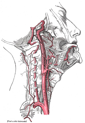
- Exception: “The 1 punch homicide” resulting in laceration of a vertebral artery as it enters the base of the skull
- Primary Traumatic Lesions of the Brain
- Penetrating injuries
- Bone fragments can act as secondary missiles
- Contusions
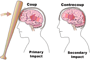
- Coup/contrecoup phenomenon – based on status of head (moving versus stationary) and impacting force (moving or stationary)
- Coup contusion is directly beneath the point of applied force
- Contrecoup contusion is located 180 degrees from the point of applied force
- Contusion tears in infants – typically at the interface of grey and white matter. Comparable to so-called “intermediate contusions” associated with
- diffuse axonal injury seen in adult brains.
- Fracture contusions – in-bending of fractured edges of skull bones impact the underlying brain
- Herniation Contusion – a secondary lesion produced by sustained pressure on the cerebellar tonsils or uncal gyri by brain swelling
- Distant contusions – contusion arising from the concussive wave of force produced by rapidly penetrating trauma, e.g. gunshot wound.
- Secondary Lesions
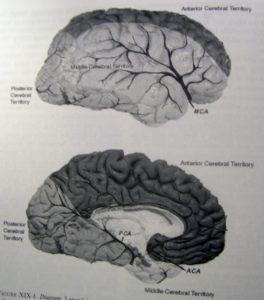
- Necrosis due to compression of a single artery, e.g. posterior cerebral compressed by herniated hippocampus
- Border zone necrosis – results from sustained hypotension with compromised perfusion front end arteries
- Duret hemorrhages – midline hemorrhage of the midbrain and pons due to sustained brain swelling
- “Respirator brain”
- Global necrosis
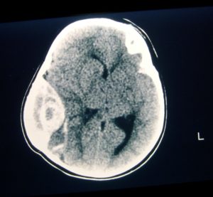
- Ischemic – inadequate perfusion due to profound brain swelling
- Hemorrhagic – typically the result of dural sinus thrombosis